Iris Galerie is hard to ignore. Perched at the top of Halkett Street, the display windows stare into your soul. It’s giving minor discomfort mixed with a prevailing feeling of awe. ‘Do my eyes really look like that?’ you wonder, staring into electric blue orbs that look ready to fuel a nuclear power plant, or deep brown craters that resemble a birds eye view of a volcano.
For those who venture in, the fascination only increases. Either Ivo – the director – or Dan – the photographer – walks you through the process of capturing your own iris, advising you to check out small details otherwise unnoticed through a magnified mirror. Those who enjoy flicking through a good coffee table book will be directed to Vision: Master Minds of Our Time by Francis Giacobetti – a stunning compilation of portraits of famous visionaries of our time, alongside detailed photos of their irises. The result is dazzling – and founder Emeric Wehbeh set up Iris Galerie so that the general public could embark on self discovery of the same fashion.
You may be thinking, ‘what’s special about my eyes?’ Or, ‘I’m not Nelson Mandela, the Dalai Lama or Stephen Hawking, why would anyone care?’ Maybe the only people who’ve ever complimented them have been disingenuous, fleeting lovers. I went to try out the experience fully aware that my irises were completely devoid of interesting details such as heterochromia, nevi or the stem cells of an absorbed twin in the womb (see above). I still found it fascinating. Ivo and Dan enlightened me on each of the minute details photographed, explaining the intricacies of how my eyes work, and became the way they are. If nothing else, it’s an experience.
Bev Corston-Petrie
This eye is so rare that it is estimated less than one in five million people share her condition. She has fetus in fetu (FIF), an uncommon developmental anomaly where a malformed, parasitic twin is found within the body of its host twin. Whilst typically found in the abdomen – hence the, “I ate my twin in the womb,” comment – FIF can occur in other locations, including the intracranial space. Bev is one of a tiny handful of people to carry her twin as stem cells in her eye.
Louisa Newman
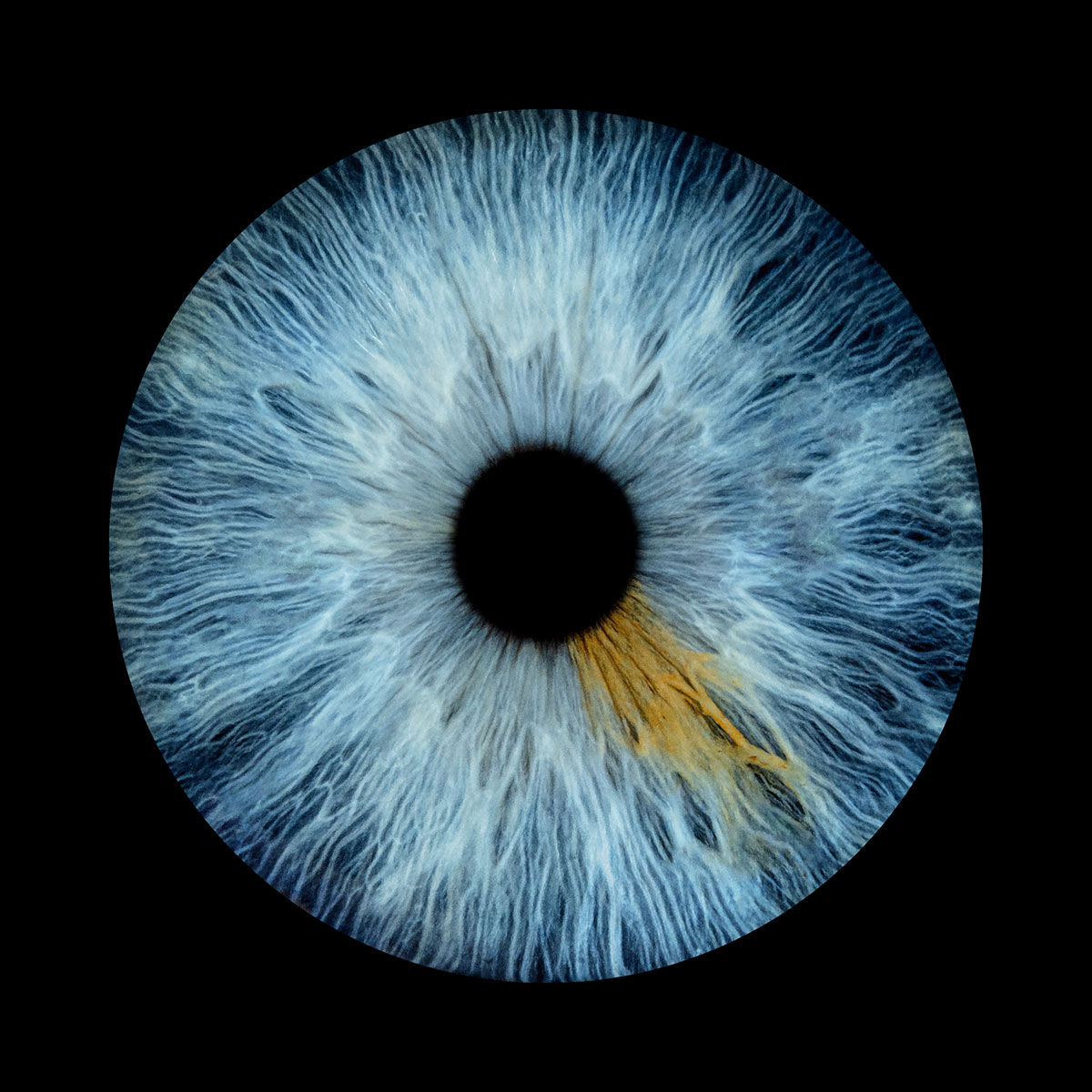
Melanin can take different forms within the iris – here, several concentrated spots called nevi combine with an amber watercolour wash that tops the blue collagen strands.
Myles Dyer
An example of partial or sectorial heterochromia – a condition where a segment of the iris has a different pigmentation.
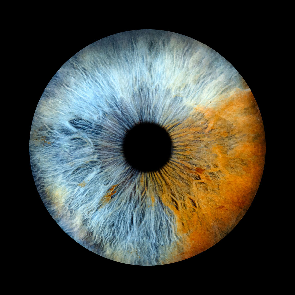
Raymond Douieb
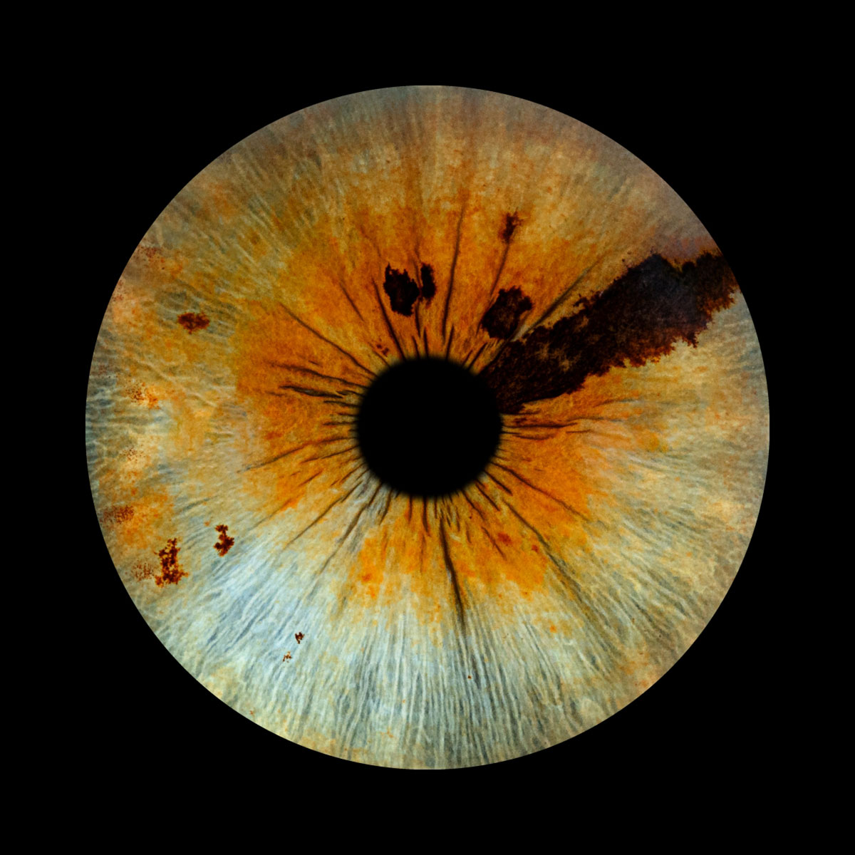
This pair of eyes combines both full and partial heterochromia, with two irises of completely different colours – the lighter showing a sectoral stripe and several nevi. The darker iris is a great example of how an increase of melanin in the eyes doesn’t just impact its colour, but its texture as well.
Sam George
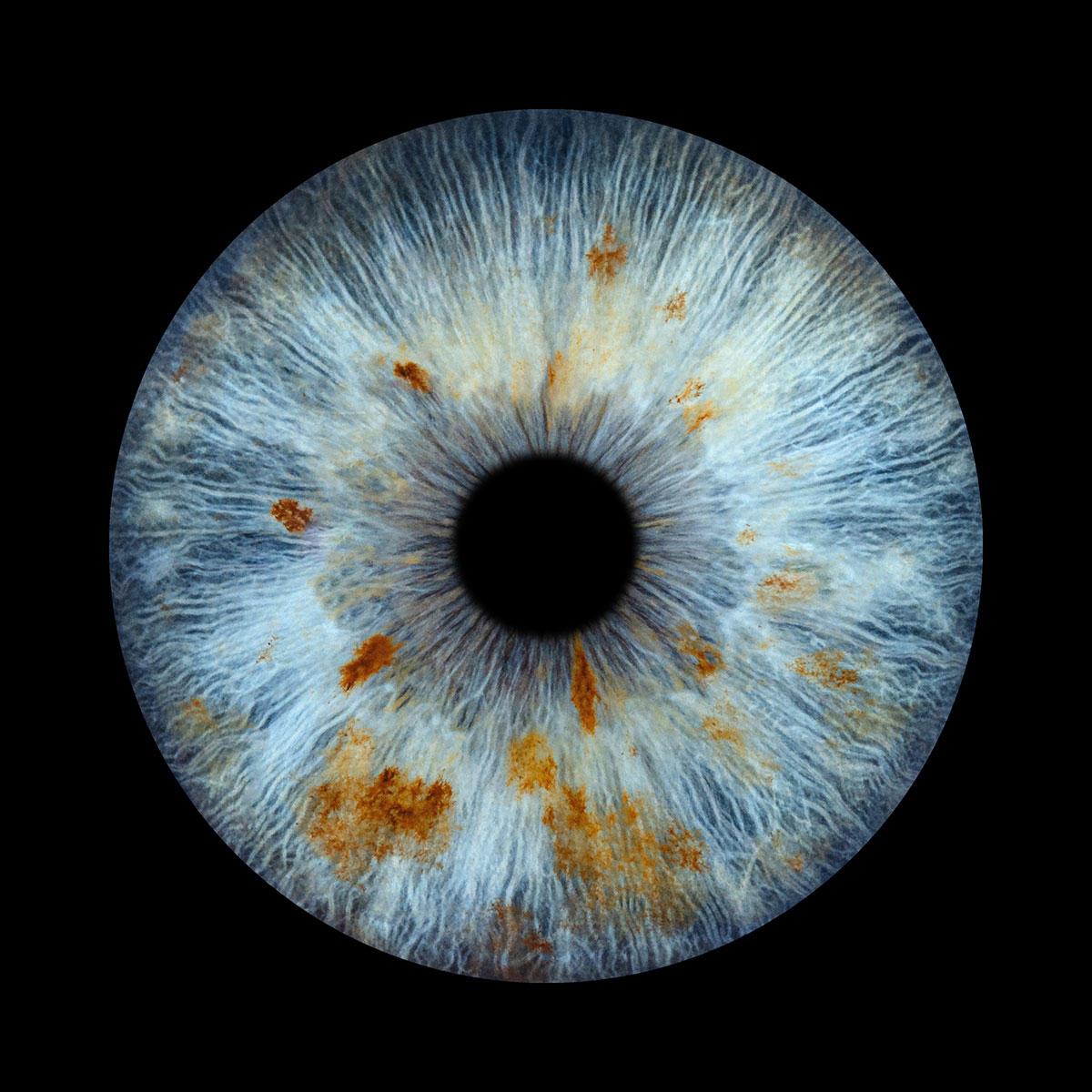
The freckle-like parts of this iris are known as Iris Nevi – caused by localised areas of melanin-producing cells which increase iris pigmentation. A nevus is a darker spot on the iris, similar to how freckles are formed on the skin.
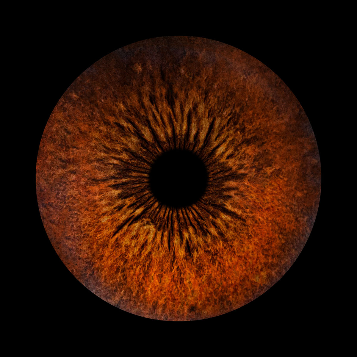
Connor Spence
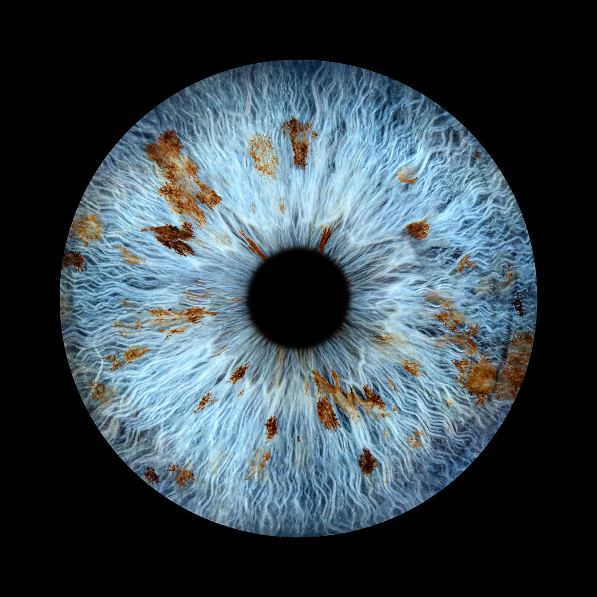
Iris Nevi caused by localised areas of melanin-producing cells which increase iris pigmentation. A nevus is a darker spot on the iris, similar to how freckles are formed on the skin. It’s usually more noticeable in lighter eyes, however can still be seen in some darker irises as well.
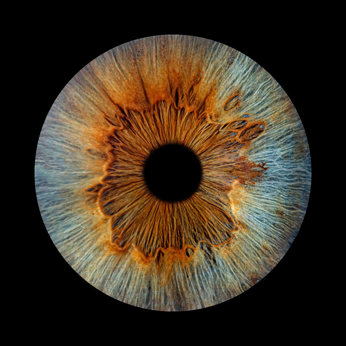
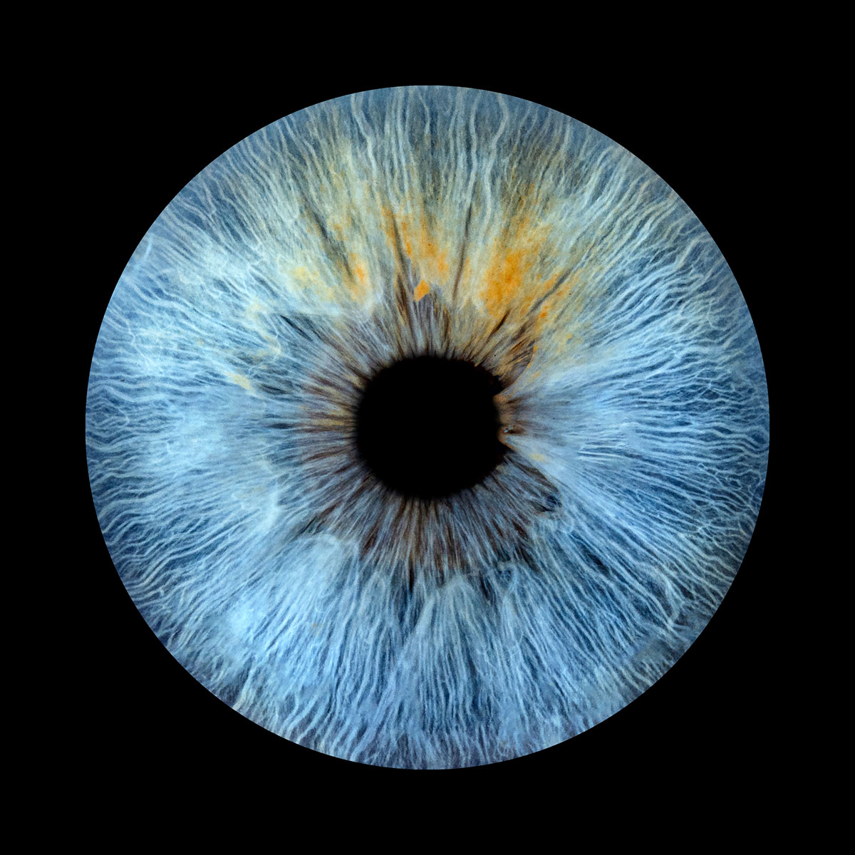
Sarah Brewster

An example of central heterochromia – a type of partial heterochromia where the centre of the iris is a different colour to the outside of the iris.
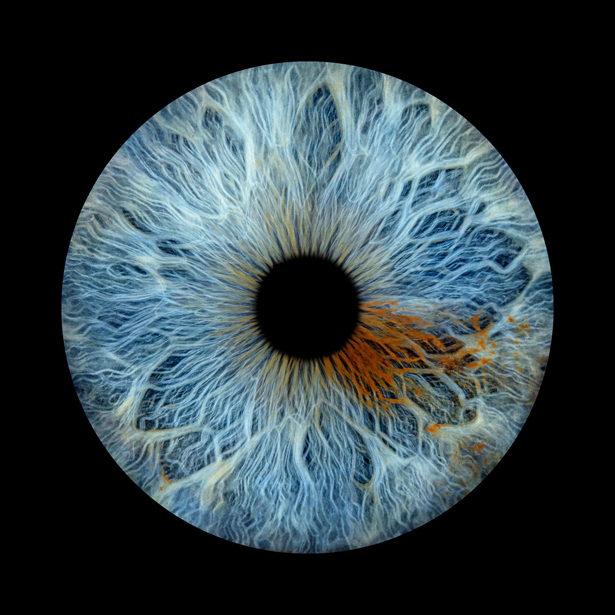
Shaun East
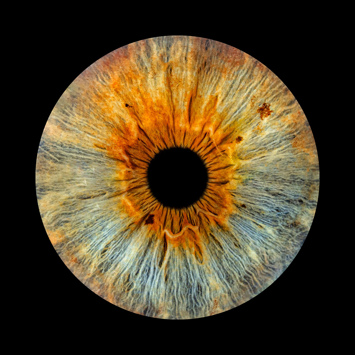
Another example of central heterochromia, but this time with the addition of nevi. They are both rare conditions – the former is responsible for the different colour at the centre of the iris, whilst the latter is responsible for the darker spots around the edge of the central “flower”.
Sophie Mercier
A great example of partial/sectoral heterochromia, showing how melanin doesn’t just change the colour of the iris, but also the texture.
Stacey Sarre
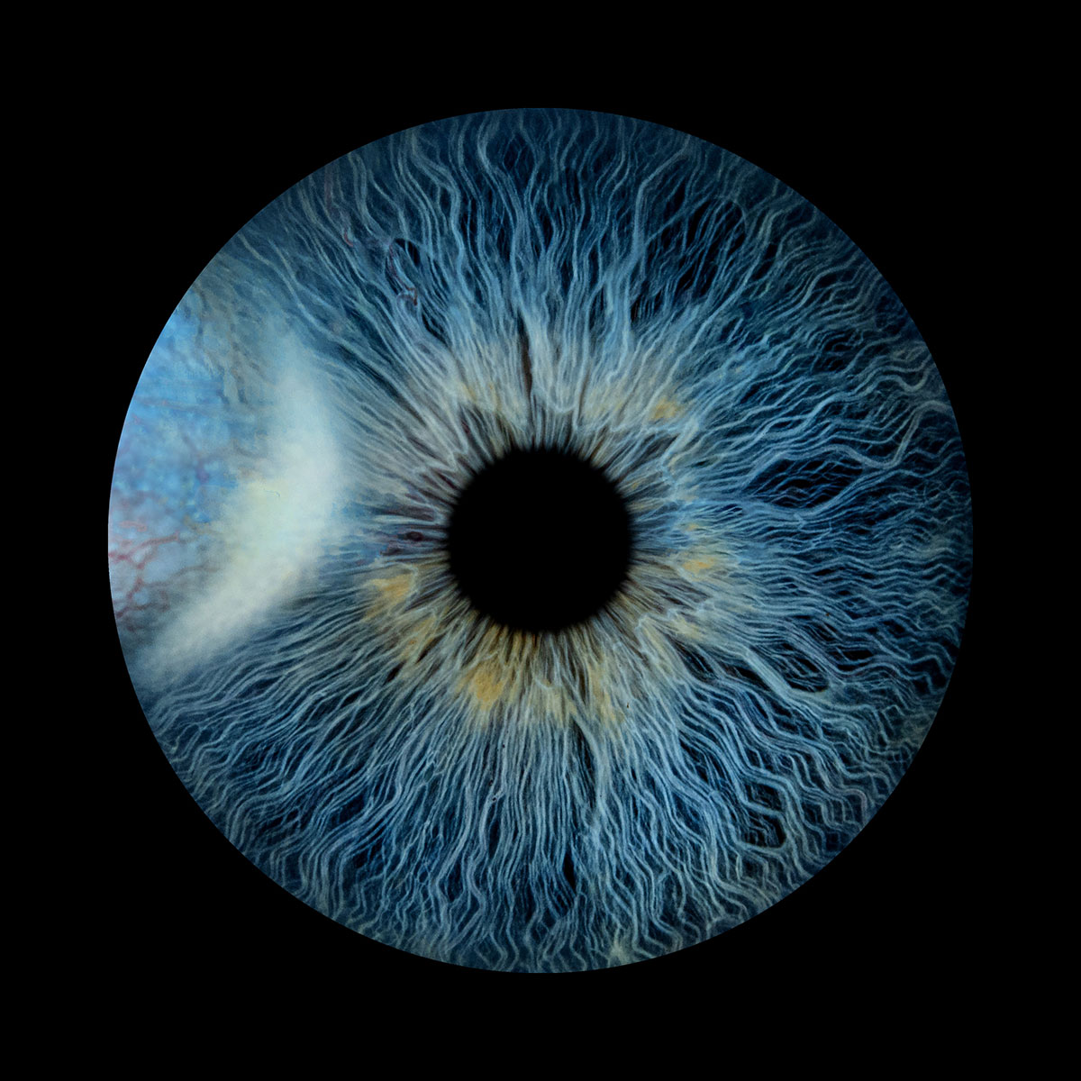
Here the tissue from the iris covers a part of the pupil – a congenital condition called persistent pupillary membrane. Most commonly seen in babies, PPM strands usually disappear within the first year, however can sometimes persist if it becomes attached to the lens or cornea.
Emma Gray
Another example of partial/sectoral heterochromia, combined with several large Crypts (places where the blue collagen strands have fused together for extra strength when opening the pupil, creating holes in the top layer of collagen and making it possible to see the layers underneath).
David Nuth
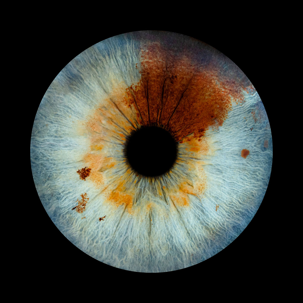
A very rare combination of sectoral and central heterochromia, including several Nevi around the lighter sections of the iris.